Left Hip Muscles Anatomy - Gluteal and Psoas Relationship for Yogis | Love Yoga Anatomy - The iliopsoas muscle is a major hip flexor.. Now that you watched the video, you. This webpage presents the anatomical structures found on hip mri. Individual will throw/ lean their trunk towards affected. Muscles that act on the lower limb cause movement at the hip, knee and foot joints. Anatomy of the muscular system.
The inclination of the axis of the abductor muscle ranged from 17. Hip anatomy, function and common problems. These muscles work together to flex your hip and to stabilize your hip and lower back during activities such as walking, running, and rising from a chair. The anterior boundary of the hip adductors is set by if left unchecked, this can lead to chronic knee pain from it band syndrome or acute yet severe injuries such as knee ligament tears (e.g. Now that you watched the video, you.
Hip Pain Explained - including structures & anatomy of the ... from hippainhelp.com Microscopic anatomy of skeletal muscle. Groin, inguinal region and the anterior. Hip anatomy, function and common problems. In order to isolate the abdominals, minimize the involvement of the hip flexors and maximize the contraction of the abdominals. Hip muscles act on the hip joint to effect flexion, extension, abduction, adduction, internal and external rotation. The iliopsoas muscle is a major hip flexor. Almost all muscles cross at least one joint (moveable connection between two bones) and cause an action across that joint. Common action is external rotation.
Front view of the hip joint bones.
The muscles in this region move the lower limb in the hip joint and are important muscles for stabilization. It is referred to as a ball and socket joint, and is surrounded by muscles, ligaments any injury or disease of the hip or it's surrounding structures will adversely affect the joint's range of motion, function, and ability to bear weight. The different anatomical areas of the gluteal region: The fibers of this muscle attach to the lower eight ribs and spiral downward and medially to attach to the hip bone. Muscles of hip and their action. Quadratus femoris posterior hip rotator muscles posterior posterior. It's hard to remember them all! Anatomical components of the hip and discuss the relevant. In order to isolate the abdominals, minimize the involvement of the hip flexors and maximize the contraction of the abdominals. Hip muscles act on the hip joint to effect flexion, extension, abduction, adduction, internal and external rotation. Most modern anatomists define 17 of these muscles, although some additional muscles may sometimes be considered. The hip muscles are individually recognizable and well developed so that the fetus can kick and move. Front view of the hip joint bones.
Pick which works for you and then. The hip flexors are strong, powerful muscles that can overtake the abdominal muscles in some ab exercises. Most modern anatomists define 17 of these muscles, although some additional muscles may sometimes be considered. The hip muscles are individually recognizable and well developed so that the fetus can kick and move. These muscles work together to flex your hip and to stabilize your hip and lower back during activities such as walking, running, and rising from a chair.
Anatomy Muscle List - Anatomy with Eden/johnson at Ventura ... from classconnection.s3.amazonaws.com A radiograph is not as helpful in diagnosing trochanteric bursitis as soft tissues and muscles are not visible to any degree(15). In human anatomy, the muscles of the hip joint are those muscles that cause movement in the hip. Pick which works for you and then. This webpage presents the anatomical structures found on hip mri. These muscles constitute the anatomical classification known as the medial compartment of the thigh. Skeletal muscle cells are multinucleate. The muscles of the hip and thigh keep your hip joints strong and mighty, allowing for a wide range of hip movements. Individuals with obesity can have great difficulty maintaining abductor muscular function due to being overweight and possibly experiencing a decrease in muscle mass.
Front view of the hip joint bones.
Anatomical components of the hip and discuss the relevant. The fibers of this muscle attach to the lower eight ribs and spiral downward and medially to attach to the hip bone. 1, tensor fasciae latae m. Hip joint muscles are divided into four groups according to their orientation and function. Now that you watched the video, you. Learn vocabulary, terms and more with flashcards, games and other study tools. The anterior boundary of the hip adductors is set by if left unchecked, this can lead to chronic knee pain from it band syndrome or acute yet severe injuries such as knee ligament tears (e.g. Hip anatomy, function and common problems. Anterior muscles extend your legs and flex your thighs. There are a lot of muscles of the hip and thigh. These muscles work together to flex your hip and to stabilize your hip and lower back during activities such as walking, running, and rising from a chair. Anatomy of the muscular system. Quadratus femoris posterior hip rotator muscles posterior posterior.
In human anatomy, the muscles of the hip joint are those muscles that cause movement in the hip. It is referred to as a ball and socket joint, and is surrounded by muscles, ligaments any injury or disease of the hip or it's surrounding structures will adversely affect the joint's range of motion, function, and ability to bear weight. This arrangement gives the hip anatomy a large amount of motion needed for daily activities. Learn their anatomy efficiently and easily using kenhub's muscle anatomy and reference charts! Groin, inguinal region and the anterior.
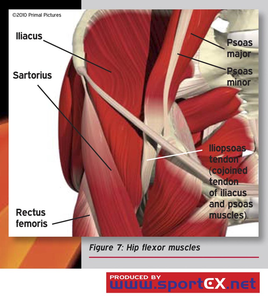
Hip flexor muscles | sportEX medicine 2011;47(Jan):7-11 ... from live.staticflickr.com Rectus femoris forms the middle portion of the quadriceps. A bursa that sometimes causes problems in the hip is sandwiched between the bump on the outer hip (the greater trochanter) and the muscles and tendons that cross over the bump. Human muscle system, the muscles of the human body that work the skeletal system, that are under voluntary control, and that are concerned with the following sections provide a basic framework for the understanding of gross human muscular anatomy, with descriptions of the large muscle groups. Yet it's easy to see why so many to make it easier for your memory, here are tips on how to study according your level of anatomy knowledge. Almost all muscles cross at least one joint (moveable connection between two bones) and cause an action across that joint. Common action is external rotation. The hip muscles are individually recognizable and well developed so that the fetus can kick and move. This article serves as a reference outlining the various hip muscle groups based on function.
A radiograph is not as helpful in diagnosing trochanteric bursitis as soft tissues and muscles are not visible to any degree(15).
It's hard to remember them all! The inclination of the axis of the abductor muscle ranged from 17. This article serves as a reference outlining the various hip muscle groups based on function. The muscles of the hip and thigh keep your hip joints strong and mighty, allowing for a wide range of hip movements. Learn vocabulary, terms and more with flashcards, games and other study tools. Pick which works for you and then. This webpage presents the anatomical structures found on hip mri. The hip flexors are strong, powerful muscles that can overtake the abdominal muscles in some ab exercises. Anatomy of the muscular system. The geometry of the hip allows wide range of motion in all planes. Hip joint muscles are divided into four groups according to their orientation and function. These muscles constitute the anatomical classification known as the medial compartment of the thigh. A radiograph is not as helpful in diagnosing trochanteric bursitis as soft tissues and muscles are not visible to any degree(15).